Movie Gallery
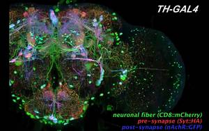
3D reconstruction of the neurons in the brain: Three different reporter transgenes are expressed in the dopaminergic neurones of the fly brain to visualise cell bodies and neuronal fibres (Green), presynaptic sites (Red), and postsynaptic sites (Blue). The three-color image nicely shows preferential distribution of output and input sites. Fluorescent serial section images obtained with a confocal laser scanning microscope are 3D-reconstructed using imaging software FluoRender. (Data by Jun Tanimura). Download movie here.
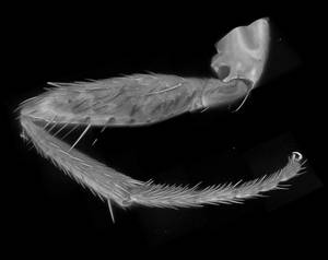
3D reconstruction of an adult leg: Insect cuticle emits autofluorescence, making it possible to make reconstruction images that look like scanning electron microscopy (SEM) images. To obtain a high-resolution image that does not fit in the imaging area of a high-magnification objective lens, multiple tiles of serial section images, each covering only a small part of the sample, were taken and concatenated. (Data by Eva Berg).
Download movie here.
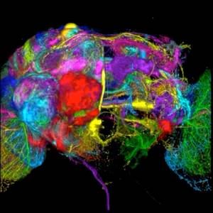
Clonal unit architecture of the brain:
Progeny of single neuronal stem cells (called neuroblasts) are labelled by inducing chromosome recombination during early development. Projection pattern of each clone is analysed in the adult brain, and images from different samples are overlaid to a common template brain to show spatial relationship among the clonal units. About 100 clones (more than 90% of the entire clones) on the left side of the brain are presented. Because about half of the clones contain neurones that project also to the other side of the brain, the right brain half is also labelled. The clonal unit serves as a developmental unit of brain composition. (Data by Masayoshi Ito).
Download movie here.
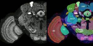
Definition of the brain regions (neuropils)
Names and boundaries of many parts of the insect brain have not been defined definitely, making it difficult to annotate locations in the brain and projection targets of neurones. To address this issue the Insect Brain Name Working Group, a consortium of 14 labs in the world, discussed and established a nomenclature system of brain structures and clearly defined brain regions. (Data by Kazunori Shinomiya). Download movie here.
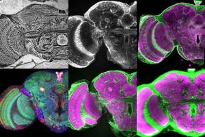
Frontal serial section images of the brain
Different staining methods reveal different structural properties of the brain. The information is used fordeliniating reasonable boundaries in the brain as shown above.
(Data by Kazunori Shinomiya) Download movie here.
- Reduced silver stain (right-top): Silver stain visualises membranes of neuronal fibres.
- gTricolorh labelling" (elav-GAL4 > UAS-DsRed, UAS-n-syb-GFP, UAS-Rdl-HA) (right bottom): Three reporters are expressed simultaneously using neuron-specific elav-GAL4 expression driver. Cytoplasmic DsRed (Red) visualises synaptic and non-synaptic fibres; synaptic vesicle-targeted n-syb-GFP (green) visualises the distribution of the presynaptic sites, similar to that obtained by nc82 antibody; and GABA receptor-targeted Rdl-HA signal (Blue) indicates postsynaptic sites and cell bodies.
- repo-GAL4 > UAS-GFP (middle top): GFP is expressed using glial specific repo-GAL4 expression driver. Glial processes form bounding sheaths surrounding many (but not all) neuropils.
- nc82 (anti Bruchpilot) antibody + repo-GAL4 > UAS-GFP (middle bottom) The nc82 antibody (Purple) labels neuropils according to the density of an active synaptic zone-specific protein Bruchpilot. repo-GAL4 > UAS-GFP (Green) visualises glial processes.
- anti-DLG (discs large) antibody + repo-GAL4 > UAS-GFP (right top): Anti DLG labels membranes of the neuronal cell bodies, fibres and synapses by detecting DLG proteins required for septate junction structure. Unlike nc82, anti-DLG also labels fibre structures within neuropils. repo-GAL4 > UAS-GFP (Green) visualises glial processes.
- anti-beta-Tubulin antibody + repo-GAL4 > UAS-GFP (right bottom): Anti-beta-Tubulin (Purple) visualises fibrous structures of both neurons and glial cells. repo-GAL4 > UAS-GFP (Green) visualises glial processes.
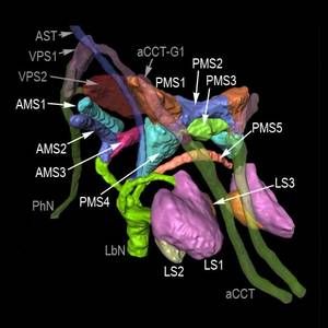
Structure of the Primary Gustatory Centre (PGC)
Axons deriving from the gustatory sensory neurons on the mouth tip project to the bottom part of the brain. They form complex branches to terminate in four major zones: ventral pharyngeal sensory centre (VPS), anterior maxillary sensory centre (AMS), posterior maxillary sensory centre (PMS) and labial sensory centre (LS). In addition gustaroy sensory neuron aoxns arising from the tips of the legs project to the region posterior to the PMS. (Data by Takaaki Miyazaki).
Download movie here.
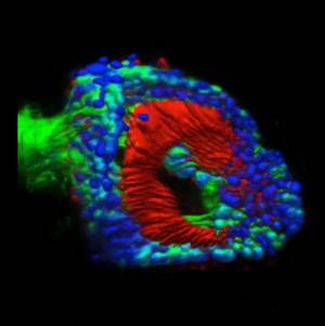
Structure of the Auditory Sense Organ (The Johnston Organ: JO)
The insect auditory sense organ that resides in the second antennal segment. About 480 tension-detecting JO Neurons are aligned like a cup, from where thin processes extend to the base of the third antennal segment. (Green: cell bodies and axons of the JO Neurons, Blue: nuclei of neurons, Red: cilia of the JO Neurons) (Data by Azusa Kamikouchi).
Download movie here.
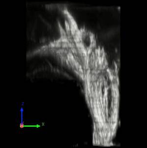
Axon terminals of the primary auditory centre
The Johnston Organ neurons project to the region called the antennal mechanosensory and motor centre (AMMC), which resides posterior to the antennal lobe (the primary olfactory centre). A thick axon bundle from the JO neurons divides gradually into smaller branches to form a complex structure. The primary auditory centre is devised into five zones and 19 subdivisions, and each JO neuron projects to one of the five zones depending on the location in the Johnston Organ. Different zones serve for the detection of different aspects of the antennal movement such as vibration for sound detection and deflection for gravity detection. (Data by Azusa Kamikouchi).
Download movie here.
Image archive of classic wall-hanging posters for zoology student courses
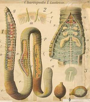
Before the age of PowerPoint, large wall-hanging posters with hand-drawn images of animals were used extensively for zoology student courses. To archive execelent but very old posters for digitial use, in total 220 posters (up to 140cm x 180cm) that have been used in the Universiity of Cologne were digitised as 150-dpi jpeg files (30-70MB each) in 2007. High-quality poster print and presentation will be possible with these files. An overview of the collection is shown by clicking the above image. The titles and abbreviations used in these posters are available here as a Word-format file.
If interested please contact Dr. Klaus Herrmann (k.herrmann(at)uni-koeln.de).